
Monkeys With Rett Pay More Attention to Stronger Social Visual Stimuli, Study Finds
Written by |
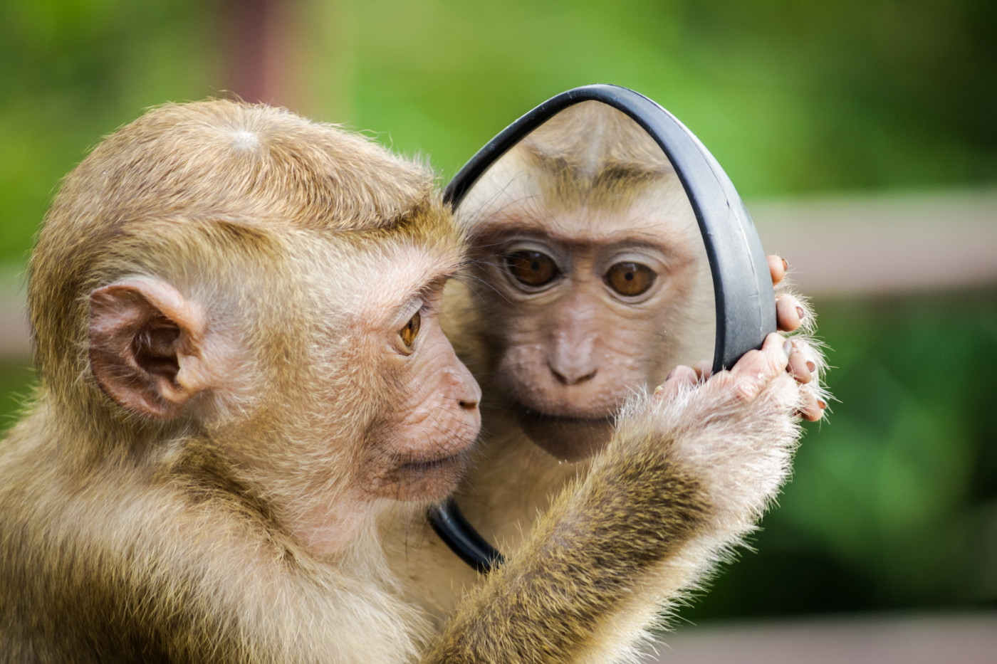


Monkeys with Rett syndrome pay more attention to stronger social visual stimuli than healthy animals do, an eye-tracking study has found.
The findings, “Social-Valence-Related Increased Attention in Rett Syndrome Cynomolgus Monkeys: An Eye-Tracking Study,” were published in Autism Research.
Rett syndrome is a rare genetic disorder characterized by developmental and intellectual disabilities. The condition is caused by mutations in the MECP2 gene — located on the X chromosome — that provides instructions to make a protein called MeCP2. This protein is responsible for maintaining synapses, which are the junctions between nerve cells that allow them to communicate.
Despite having severe motor and speech impairments, people with Rett syndrome normally retain their ability to communicate and express their feelings through visual cues.
“Clinical eye-tracking studies have furthermore revealed unique attentional features in [Rett] patients, such as attentional preference to salient, novel, and socially weighted stimuli, indicating the procedure as an effective avenue to quantify the visual cognitive phenotypes [features] associated with the syndrome,” the investigators said.
In this study, a group of researchers from the Kunming University of Science and Technology in China set out to evaluate different parameters of visual attention — the period of time spent looking at an image — in monkeys with Rett syndrome. To do so, they performed several behavioral tests in which they followed the animals’ eye movements. These included:
- The social valence comparison (SVC) test, in which investigators showed animals two images of the same monkey’s face. One image was in profile, depicting mild stress; the other was staring directly, depicting fear. The researchers assessed how much attention the monkeys paid to visual cues representing different types of social stimuli.
- The visual paired comparison (VPC) test, showing the monkeys two identical images and then replacing one with a new image The investigators evaluated whether the animals preferred to spend more time observing the new image.
- The social recognition memory (SRM) test, in which researchers showed the monkeys a series of images. Investigators first showed the animals an image of a monkey, followed by several images of the same animal taken at different angles. Finally the monkeys were shown a new set of images from a different animal. The goal was to see if the animals were able to recall the monkey they had seen previously.
In parallel with behavioral tests, the investigators also performed magnetic resonance imaging (MRI) scans to look for possible brain alterations in the monkeys. The team also collected blood and cerebrospinal fluid samples from the animals to measure the levels of neurotransmitters, in an effort to pinpoint the mechanisms that could be associated with the animals’ behavior.
Cerebrospinal fluid (CSF) is the liquid that surrounds the brain and spinal cord. Neurotransmitters are chemical substances that allow communication between nerve cells.
Results showed that, in the SVC test, monkeys with Rett syndrome spent much more time looking at stare faces than profile faces. In comparison, the healthy animals spent approximately the same amount of time looking at both types of images.
“[This] result suggested attentional preference to the more salient social stimuli in RTT monkeys, consistent with previous clinical findings,” the researchers said.
In the VPC and SRM tests, all animals showed preference for the new images they had never seen before. That indicates that short-term memory remained unaffected in the monkeys with Rett syndrome.
In addition, researchers found that in the VPC and SRM tests, the monkeys with Rett syndrome tended to spend more time looking at images of monkey faces than other animal faces, compared with controls.
MRI brain scans revealed that animals with Rett syndrome had a significant reduction, compared with healthy animals, in the size of the occipital gyrus, a region of the brain involved in the processing of visual cues, on both sides of the brain.
“The reduced bilateral occipital gyrus of the RTT monkeys could induce or partially contribute to the increased attention findings,” the investigators said. “However, the specific structural and functional changes of the visual pathways/cortex associated [with] the syndrome … remain unclear and further investigations are required to clarify the issue.”
No significant changes were found in the levels of neurotransmitters present in the blood or in the CSF of diseased animals compared with controls.
“The present eye-tracking study found social-valence-related increased attention in the MECP2 mutant RTT monkeys, supplementing the cognitive phenotypes associated with the syndrome. Without revealing sufficient explanatory brain and neurotransmitter abnormalities, further investigations from broader perspectives are required to uncover the underlying neurobiological mechanisms,” they said.

Leave a comment
Fill in the required fields to post. Your email address will not be published.